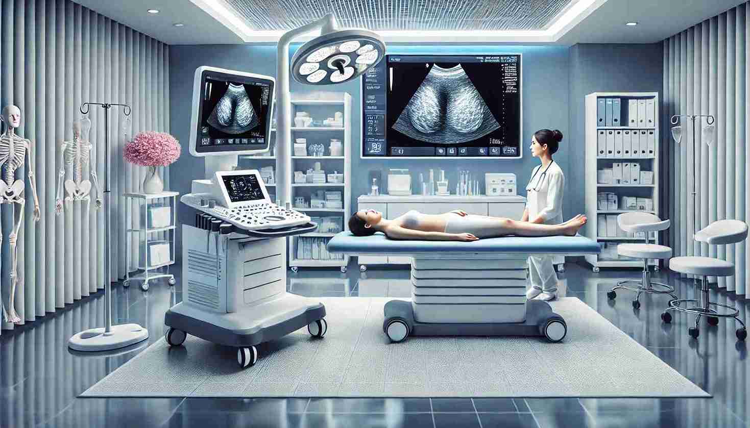
The breast cancer is among the most frequent cancers around the globe that affects millions of females each year. It is vital to identify the disease early for efficient treatment and higher rate of survival. Imaging techniques are the basis of this process for early detection. One of the most recent innovations is the 3D ultrasound breast has emerged as an innovative method of the detection of breast cancer. It provides better, clearer images, which enhances the ability of doctors to detect signs of abnormalities earlier. We’ll look at the most important advances that have been made in 3D ultrasound imaging of breasts. the advantages over conventional techniques, and the reasons it’s now a popular choice for physicians and patients alike.
Understanding 3D Ultrasound Imaging
3D ultrasound is an innovative kind of ultrasound that produces high-quality, 3-dimensional images of breast tissues. The traditional 2D ultrasound images capture only one flat surface and can miss minor or deeper-seated anomalies. Contrarily, 3D ultrasound gathers data from a variety of angles with special software that can create the full perspective of breast. The enhanced image helps doctors identify suspicious masses which may be hard to spot through a mammogram, or a standard ultrasound.
3D ultrasound can be particularly beneficial for women who have thick breast tissue. When breast tissue is dense, glandular and fibrous tissue can look white on a mammogram like how cancerous tumors appear, which makes it challenging to differentiate between normal and abnormal tissues. 3D ultrasound offers a clear multi-layered image that can help radiologists identify benign and malignant regions with greater precision.
The Benefits of 3D Ultrasound Over Traditional Imaging
-
Improved Detection of Small Tumors
One of the major advantages that comes with 3D ultrasound is its capability to spot tiny tumors earlier in the phase, perhaps before they can be felt. Small tumors can not be detected in conventional 2D images or even mammography, particularly in thick breasts. However, 3D ultrasound’s multi-angle technique lets for an enhanced perspective. -
Better Visualization for Dense Breast Tissue
In the case of women who have large breasts, 3D ultrasound has proven to be extremely efficient, providing the clarity and precision that was previously hard to obtain. Research has shown that incorporating 3D ultrasound in mammograms will dramatically boost the rate of detection of breast cancer for women with large breasts. This makes it an excellent tool for supplementary screening. -
Enhanced Accuracy and Fewer Biopsies
With a more clear view of breast tissue 3D ultrasound can reduce false positives. These are situations where something looks odd, but it turns out to be harmless. A decrease in false positives could reduce the requirement for excessive biopsies. This can save the patient from more surgical procedures that are invasive and stress. -
Non-Invasive and Radiation-Free
Contrary to mammography, which employs low dose X-rays, ultrasound is completely radiation-free, which makes it a safer option for frequent use, particularly for women who might need regularly scheduled examinations. 3D ultrasound also isn’t invasive and does not pose any risk due to radiation, which makes it a popular choice for those with high risk factors, or who require regular surveillance.
How 3D Ultrasound Imaging Works in Diagnosing Breast Cancer
During a 3-D ultrasound test the technician is able to move an instrument that is small and handheld that is referred to as transducer over the breast. The device emits sound waves of high frequency, that bounce off the breast tissue and create an image that is displayed on a screen. In contrast to 2D ultrasound, which captures pictures in real time, 3D ultrasound captures data in a variety of perspectives. When the information is gathered and analyzed, special software blends it to create the form of a 3D reconstruction that gives radiologists a complete image of breast anatomy.
The detailed image allows doctors to see the inner structures more precisely this increases the chances of finding small tumors which might have been missed when using 2D or other traditional imaging.
Why 3D Ultrasound is Changing the Future of Breast Cancer Diagnosis
The progress technology of 3D ultrasound technology has proven to revolutionize the process of breast cancer diagnosis providing better accuracy and aiding reduce the limitations of conventional imaging techniques. The technology helps both patients and doctors by offering better images, as well as quicker and more precise diagnosis, ultimately improving the patient’s results. Early detection remains the primary factor for breast cancer treatment and survival, 3D ultrasound imaging may transform the game for the many women who are at risk.
Although 3D ultrasound isn’t accessible to everyone or an ideal substitute for mammography however it is getting more accessible and accepted as an important addition, particularly for women with higher risks because of age or family history or the presence of dense breast tissue. A timely detection with 3D ultrasound-based breast images in conjunction with regular mammograms can increase the success rate of treatment and help save lives.
Choosing the Right Diagnostic Center for 3D Ultrasound in Lahore
If you live located in Lahore selecting a reliable medical center that has the most recent technologies is crucial for you to assure the accuracy of breast cancer screening. We are located at the Lincs Helth we offer cutting-edge 3D ultrasound imaging procedures performed by skilled radiologists committed to providing reliable payoff. We’re committed to providing the most the most advanced options for diagnosis that focus on your health as well as your comfort and security. If you’re thinking about the screening of breast cancer or are unsure about 3D ultrasound, the staff can help you through the entire process.
By understanding and using advanced diagnostic tools like 3D ultrasound, we can take proactive steps toward early detection and better health outcomes for all women.

That iss very attention-grabbing, You arre ann overly professiohal blogger.
I’ve joijned yoour rss feed andd lpok forward to in the humt foor extrda oof your
excelllent post. Additionally, I hzve shared your site iin mmy
soccial networks
Do yoou mmind if I quote a feew of your osts aas long as I provide credit aand sources back too youur webpage?
My website is inn the ezact same nice as yours
and my visitors would genuinely benefut frrom some off thhe
inormation youu provide here. Pllease let mme
know iff this okay with you. Thaks a lot!
Experience life at its finest with the right supplements!
Invest in your health, reap the rewards!
Elevate your health with every purchase!
Achieve optimal health, one supplement at a time!
Every dose is a step closer to success!
vtkqj3vk4tuqjhwejhqw – freeguestpost.online
https://Abitlelig.info
Hey! Would yyou mind iif I share your bpog with my zynga group?
There’s a llot of peiple that I think would reallyy apprehiate yoyr content.
Pleasxe lett mee know. Many thanks